Background and aims
Skin graft necrosis is a common complication in dermatologic surgery, but reliable methods for predicting its occurrence are lacking. Laser speckle contrast imaging (LSCI) is a validated tool for studying skin graft microcirculation. We aimed to analyze the relationship between perfusion, assessed intraoperatively or postoperatively with LSCI, and the extent of necrosis on day 28. In addition, we aimed to determine whether perfusion can predict the development of clinically significant necrosis, defined as necrosis affecting 20% or more of the graft.
Materials and methods
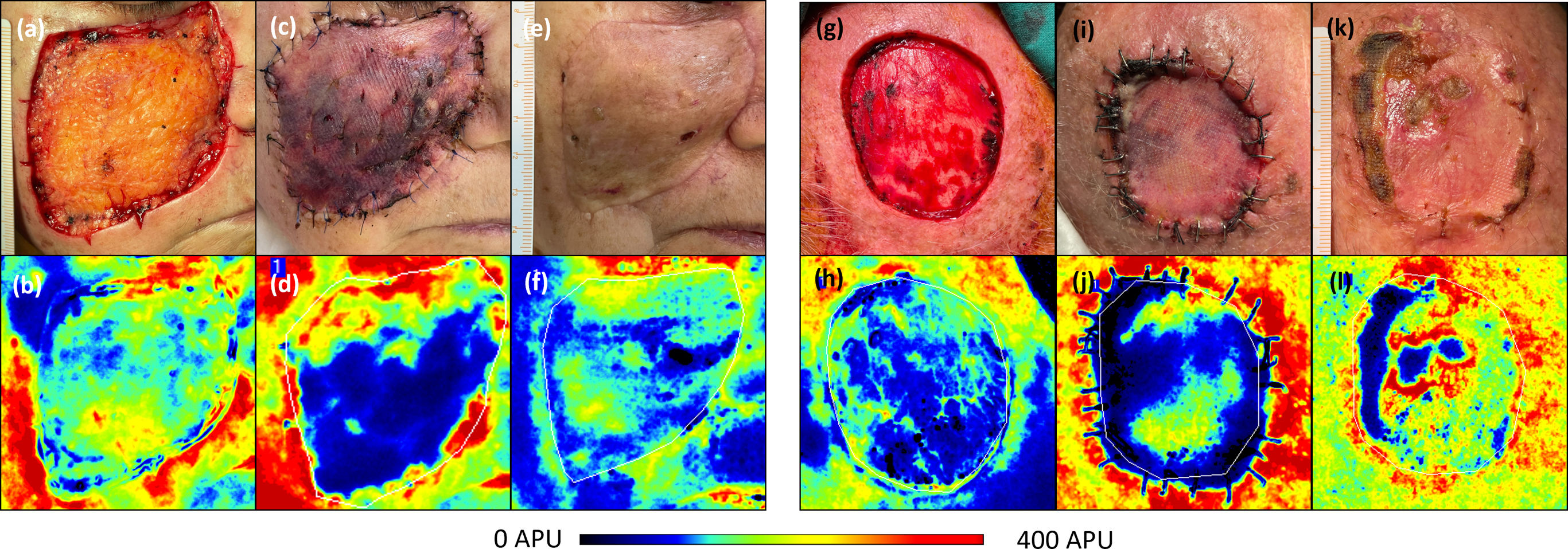
Fifty-five adults undergoing skin grafting after skin cancer excision were included. Perfusion was assessed with LSCI intraoperatively on the wound bed and on the graft surface on day 7. The extent of necrosis was determined on day 28.
Results
Graft necrosis was significantly correlated with graft bed perfusion assessed on day 0 (r=−0.86) and graft perfusion assessed on day 7 (r=−0.69). Significant linear regression models showed that intraoperative graft bed perfusion and day 7 graft perfusion explained 75% and 46% of the variability in necrosis extent, respectively. Overall, each unit increase in graft bed perfusion reduced necrosis extent by 41 percentage points, whereas each unit increase in day 7 graft perfusion reduced necrosis extent by 21 percentage points. The perfusion threshold with the highest predictive value for clinically significant necrosis was identified in the graft bed.
Conclusions
LSCI may provide valuable information as a predictive tool for necrosis through rapid, noninvasive perfusion analysis, allowing the identification of patients who are optimal candidates for skin grafting and those who may require prolonged wound care.